Structural Joint Types and Common Injuries
Objectives
Objective #1:
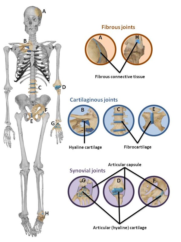
_Three joint types categorized by structure with descriptions.
-Major Examples with images of each structural type.
Objective #2:
_Traumatic Joint Injuries: Total Knee Reconstruction.
-Describe Injury:
.Three major ligaments typically damages
.Test to Diagnose
.Procedures leading up to surgery
-Surgical Procedures
-Treatment and Rehabilitation
Objective #1:
Cartilaginous Joints-
Cartilaginous joints allow very little or no movement, and are categorized by the connection between the adjoining bones made of cartilage. Cartilaginous joints are joints covered with cartilage, such as hyaline or fibrocartilage, to allow movements between bones more than fibrous joints but less than synovial joints. Cartilaginous joints are divided into two groups: primary and secondary. Primary cartilaginous joints, or synchondroses, often have cartilage, hyaline, or fibrocartilage that are converted to bone with age. Exceptions of this process are the joints holding the first rib to the manubrium of the sternum, and sternal synchondroses. Secondary cartilaginous joints, or symphyses, often are flat disks of fibrocartilage that connect bones and remain un-ossified throughout life. Examples of slightly moveable cartilaginous joints include the pubic symphysis of the pelvis and the intervertebral joints of the spinal column where the articulating bone surfaces are connected by pads, or discs, of cartilage. Examples of immovable cartilaginous joints include the hyaline cartilage epiphyseal plates of growing long bones and the cartilaginous joints between the first ribs and the sternum.
http://www.ivy-rose.co.uk/HumanBody/Skeletal/Joints/Cartilaginous-Joints.php
Fibrous Joints-
Fibrous joints are immovable or fixed joints in which movement between bones is possible. These joints are immovable because the bones are held firmly together by bundles of strong white collagen fibers. There are three types of fibrous joints: sutures, syndesmoses, and gomphoses. Sutures are immovable joints consisting of a thin layer of dense fibrous connective tissue attaching certain bones of the skull. Syndesmoses are attached by fibrous connective tissues, which are neither a suture nor a gomphosis. Gomphoses are immovable joints in the shape of a socket at which one solid structure in an individual's body is firmly attached to another. Examples of fibrous joints include the sutures between the skull, syndesmoses between certain long bones, like the tibia and fibula, and gomphoses that attach the roots of human teeth to the upper and lower jaw bones.
http://www.ivy-rose.co.uk/HumanBody/Skeletal/Joints/Fibrous-Joints.php
Synovial Joints-
Synovial joints are the most movable joints. Their surfaces are covered with hyaline cartilage and their articular cartilage is avascular, non-nervous and elastic. Synovial joints are lubricated with synovial fluid, the cartilage forms slippery surfaces for free movements. there are six types of synovial joints: plane joints, hint joints, pivot joints, condyloid or ellipsoidal joints, saddle joints, and ball-and-socket joints. Plane joints have flat articular surface. Hinge joints are cylindrical projections(condyles) that fit into concave shapes. Pivot joints have a rounded end of one bone that fits into the sleeve of bone or ligaments. Condyloid or ellipsoidal joints have an oval surface of one bone that fits into the depression of another bone. Saddle joints have both concave and convex surfaces that allow for more movement. Ball-and-socket joints have a spherical head of joe that articulates into a cup like surface of another. Examples of synovial joints include the knee joints, hip joints, shoulder joints, and elbow joints.
http://www.ivy-rose.co.uk/HumanBody/Skeletal/Joints/Synovial-Joints.php
http://faculty.stcc.edu/AandP/AP/AP1pages/Units5to9/joints/synovial.htm
http://legacy.owensboro.kctcs.edu/gcaplan/anat/notes/api%20notes%20i%20types%20of%20joints.htm
Objective #2:
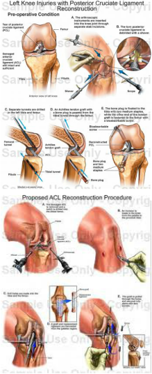
Total knee reconstruction would inquire that an individual has injured each of his/her major ligaments: the Anterior Cruciate Ligament(ACL), the Posterior Cruciate Ligament(PCL), the Lateral Collateral Ligament(LCL), and the Medial Collateral Ligament(MCL). The ACL is one of the two major ligaments in the knee. It connects the femur to the tibia in the knee. The PCL is the second major ligament that connects the femur to the tibia in the knee. The LCL connects the femur to the fibula, the smaller bone of the lower leg on the lateral(outer) side of the knee. The MCL also connects the femur to the tibia on the medial(inner) side of the knee. Doctors diagnose by first checking to see if an individuals knee is swollen. The extent and speed of joint swelling may indicate the seriousness of the injury. The doctor will also check for soreness along the collateral ligaments(where they are connected to the bone). After checking for these symptoms the doctor will carefully check for lateral looseness by moving the knee from side to side, both when the knee is bent and straight. If the injury is acute, the knee may be too sore to examine throughly. In this case, the doctor might either apply a local anesthetic to the knee or put a splint on the knee and re-examine it after eight to ten days. Procedures an individual must follow prior to surgery include rest, ice pack application(to reduce swelling), compression(from an elastic bandage or brace), elevation, and pain relievers. Before the surgery the doctor will also go through a series of questions with the patient to check for medical history, disorders, medicines being taken, possible pregnancy, etc. For sever collateral ligament tears, an individual may need surgery to attach the ligament back to the bone if it was pulled away or to the other part of the ligament if it was torn in the middle. Unfortunately, neither the ACL nor PCL are able to be repaired once they are completely torn or stretched beyond their limits. The only option is RECONSTRUCTION. In this procedure, tendons are taken from other parts of an individuals leg or a cadaver to replace the torn ligament. Ligament reconstruction for an ACL or PCL injury is complicated and involved. This surgery is not for everyone. Those who choose not to have surgery must accept the pain and risk of some permanent weakness and instability in the leg. Treatment/rehabilitation for individuals who have just recently had knee ligament reconstruction surgery include the possibility of having to wear a knee brace for the first one to four weeks. Some may even be required to be on crutches. However, most people are able to move their knee right after surgery to help prevent stiffness. An individual may also be given pain medication. Physical therapy is highly recommended to help an individual to help regain motion and strength within the knee. Therapy can last two to six months. A full return to activities and sports usually take about four to six months.
